New research instruments bring new possibilities to Binghamton
University invests in MRI scanner, HAXPES system, 3D X-ray microscope

Three major pieces of state-of-the-art research instrumentation recently debuted at Binghamton University, each with potential to contribute to advances in fields such as engineering, materials science and human health.
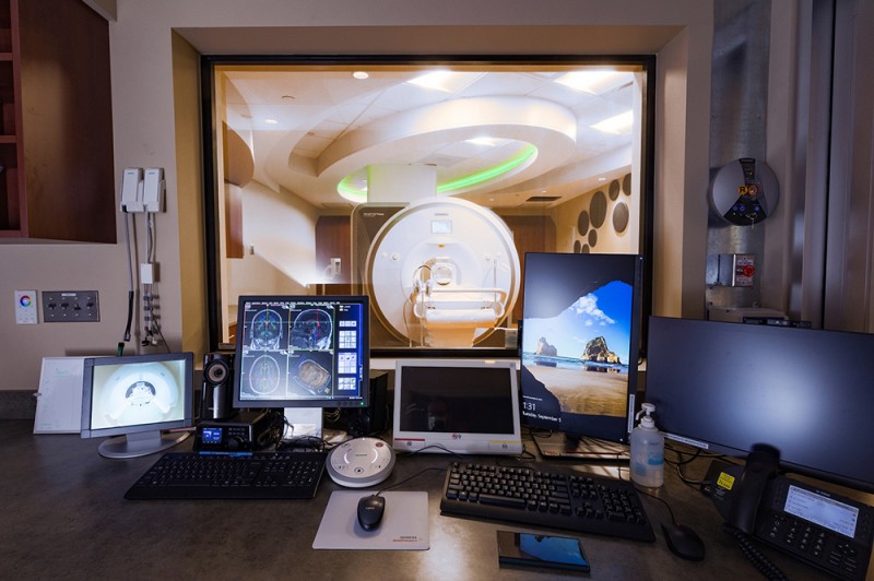
Magnetic resonance imaging (MRI)
The University purchased a $2.6 million Siemens Magnetom Prisma 3 Tesla scanner as part of a collaborative venture with United Health Services, leading to the establishment of the Binghamton Brain and Body Imaging Research Center.
The center, led by Distinguished Professor J. David Jentsch, will deepen our scientific understanding of brain disorders.
“The fundamental goal of MRI is to see what should be unseeable, what’s inside the deep recesses of your body,” Jentsch says. “But that’s kind of what makes MRI remarkable. I like to think of this as being one of the few machines that the human brain created to see and to heal itself. That really sets it apart from other kinds of healthcare and research technology.”
The project was about a decade in the making, says University President Harvey Stenger, who notes that it’s novel to embed a research tool inside a working healthcare facility.
“It’s almost impossible to think about the brain being understood like we understand a bone or a muscle,” he says, “but with the new computational artificial intelligence breakthroughs, professors like David Jentsch and future faculty, I really do think that the brain will be understood at a fundamental level so that we can predict the cause of terrible diseases and disorders like autism and Parkinson’s and Alzheimer’s. That’s the goal.”
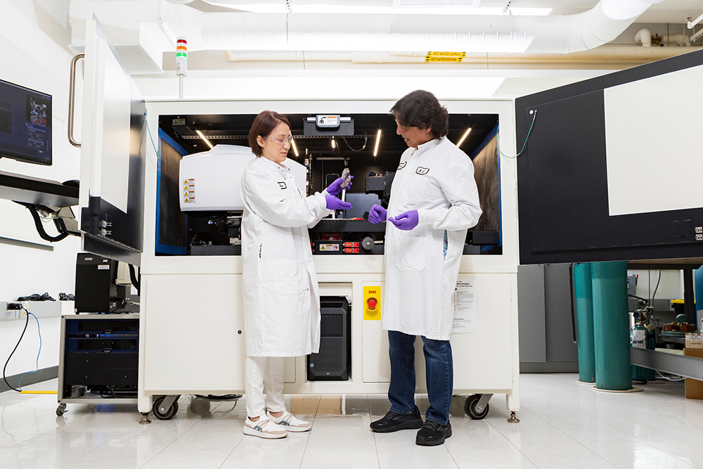
HArd X-ray Photoelectron Spectroscopy (HAXPES)
The University’s Analytical and Diagnostics Laboratory, part of the S3IP Center of Excellence, offers core facilities to the entire campus community as well as to industry partners. The lab acquired a HArd X-ray Photoelectron Spectroscopy, or HAXPES, system with support from the National Science Foundation’s Major Research Instrumentation program.
The $2 million Scienta Omicron HAXPES tool is the first of its kind in North America.
Matthew J. Wahila, chief technology officer for the center, says the lab is especially useful for scientists who want to get the “fingerprint,” or chemical composition, of a sample. For instance, the Binghamton lab has already been used by chemists to analyze cathode powders for batteries.
“It helps you understand the samples that you’re fabricating,” Wahila says. “Exact composition can be very important, especially if you’re talking about doping materials where you could be putting in a fraction of a percent of one type of atom and that can have drastic effects on the properties.”
HAXPES will enable scientists to get that kind of understanding and iterate or improve their fabrication procedures more quickly than if they had to wait for an opportunity to travel to a synchrotron to conduct an experiment.
3D X-ray microscope
The Analytical and Diagnostics Laboratory added a $1.7 million Zeiss 620 Versa Xradia instrument to its suite of tools, which are available to campus researchers and industry partners. The Xradia, a 3D X-ray microscope, is capable of tremendous magnification without destroying a sample.
Senior scientist Anju Sharma says the versatile instrument is suited to a broad range of applications; it can capture images of biological materials, whole electronic devices, 3D printed parts, batteries, rocks, fossils and more. Images in 2D are collected as a sample rotates under X-ray; powerful software reconstructs them into a 3D volume that can be virtually cross-sectioned at any location or orientation.
“It can do truly nondestructive characterization and give you very high-resolution data,” Sharma says. “You can look at the micro world inside a large-scale object.”
She says a quick scan can be conducted in an hour, while other tests may take a few hours or more. The Xradia system also enables scientists to do “4D experiments,” in which a 3D microstructure is subjected to compression, extension or changes in temperature over time.
Sharma recently worked with an industry user to examine a large circuit board that had experienced an electrical failure. The Xradia made it possible to find a tiny crack in a solder joint without destroying the board.
“With other electron microscopes, you can look at the surface,” she says. “You can’t look inside.”