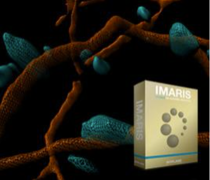
 Imaris is the world’s leading interactive microscope Image analysis software. It provides
smart detection and visualization of complex objects, tracing of neurons, blood vessels
and other filamentous structures, 3D tracking, interactive plotting, and batch analysis.
Imaris is the world’s leading interactive microscope Image analysis software. It provides
smart detection and visualization of complex objects, tracing of neurons, blood vessels
and other filamentous structures, 3D tracking, interactive plotting, and batch analysis.
The software has 4 detection models. Spots, surface, cells and filaments which can be harnessed to detect and analyze almost all biological samples, including cells, nuclei, bacteria, organs, dendritic spines, blood vessels, and many more.
- Cell analysis:
Volume, vesicles per cell, distance to membrane, morphology
- Motion analysis:
Speed, acceleration, cell division tracking, trajectory, event synchronization
- Quantification:
Count of objects, area, volume, intensity, position
- Interactions:
Distances between structures, volume overlap and contacts, 3D intensity profiling, spatial distribution, co-localization
- Batch and plots:
Image processing, measurements, interactions, group comparisons, statistical tests
- Filaments/neurons:
Length, straightness, mean diameter, branching angle, spine density, spine shape
Please refer to Imaris Homeschool to learn specific applications using the software.